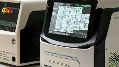
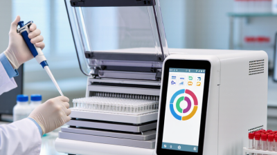
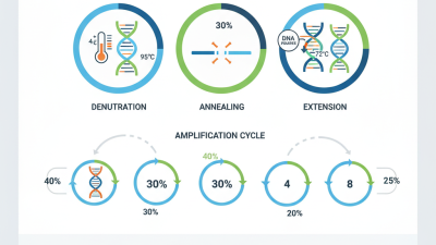
Accurate PCR results begin with disciplined preparation, not with the final fluorescent curve. Each stage affects how confidently a laboratory can detect and interpret genetic material. The pcr steps include sample preparation, reagent setup, denaturation, annealing, extension, and result analysis. Small errors can appear as major problems later. A poorly mixed sample may produce uneven concentrations. Incorrect primer design may create weak or nonspecific signals. Even a brief temperature variation can influence amplification efficiency.
Experienced laboratory teams treat every stage as a possible source of uncertainty. They use calibrated pipettes, clean workspaces, appropriate controls, and validated protocols. Negative controls help reveal contamination. Positive controls show whether the reaction worked as expected. Careful timing also matters, especially during repeated temperature cycles. The details are practical: a misplaced tube, a wet pipette tip, or a delayed transfer can change the outcome. These issues may seem minor. They are not.
Reliable PCR requires more than following a printed procedure. Scientists must understand why each condition exists and review unexpected data honestly. A late amplification signal may indicate low target levels, inhibition, or contamination. It should not be accepted automatically. No protocol is flawless. Even well-trained professionals can miss an unusual variable. That is why quality assurance, documented observations, and independent review remain essential. Understanding the pcr steps helps researchers connect technical choices with trustworthy conclusions. It also encourages better questions when results seem convincing, but the evidence is incomplete.
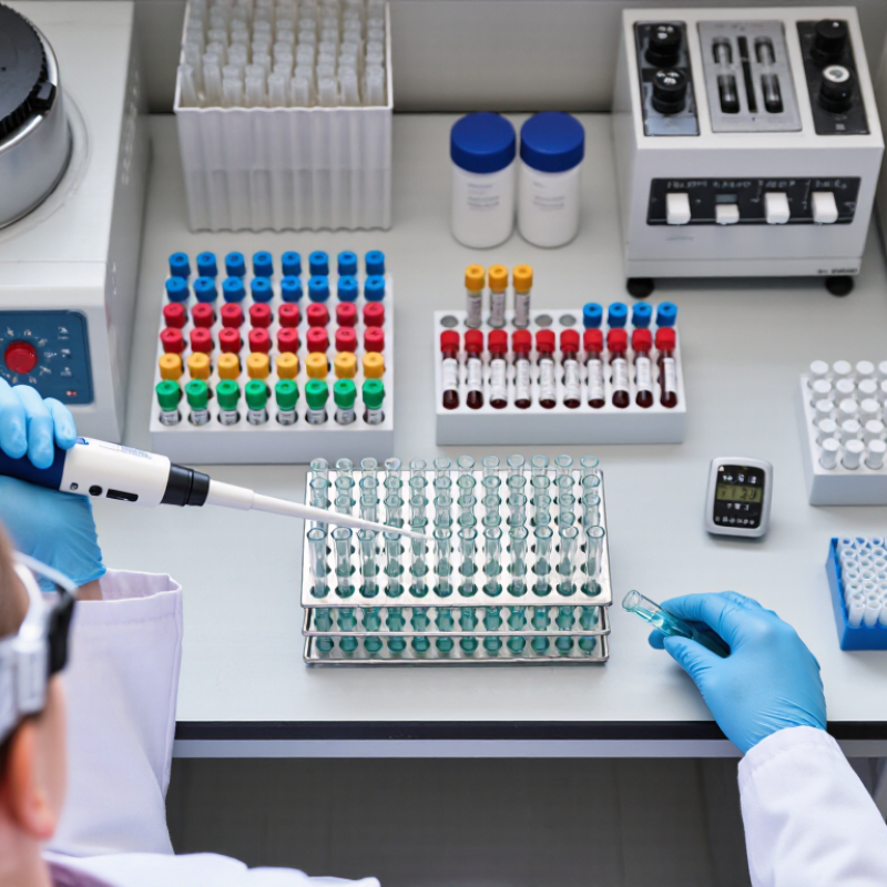
Accurate PCR begins before the first cycle. A clean tube does not guarantee a clean template. Blood proteins, salts, ethanol, and residual detergents can inhibit polymerase activity. Even a small amount of carryover may delay amplification or produce weak signals.
DNA concentration matters, but purity matters more. Many laboratories use an A260/280 ratio near 1.8 as a practical quality check for DNA. This number is useful, but it is not proof of integrity. A fragmented template can show an acceptable ratio and still perform poorly. I have seen concentration readings look excellent while amplification remained inconsistent.
The MIQE guidelines recommend documenting sample processing, nucleic acid quality, and inhibition controls. These details make results easier to evaluate and reproduce. A Nature survey of 1,576 researchers found that more than 70% had failed to reproduce another scientist’s experiments, while about half had failed to reproduce their own. Sample preparation is not the only cause, but it is an overlooked variable. The U.S. Centers for Disease Control and Prevention also identifies substances such as heparin, hemoglobin, and ethanol as potential PCR inhibitors. Careful washing, controlled elution, and proper storage can reduce these risks. Still, every workflow has weak points. Temperature changes, repeated freeze-thaw cycles, and rushed pipetting can quietly damage confidence in the final result.
Denaturation is the first decisive temperature step in PCR. Heating separates double-stranded DNA into two single strands. Each strand becomes a template for primer binding and extension. Most protocols use 94–98°C for 10–30 seconds, depending on the polymerase and amplicon. Too little heat leaves duplex regions intact. Excessive heat can reduce enzyme stability. Small settings matter.
In routine assay development, I watch the target’s melting behavior, not only the programmed temperature. GC-rich regions may need stronger or longer denaturation. The MIQE guidelines, published in Clinical Chemistry, identify 90–110% as a practical amplification-efficiency range for quantitative PCR. Incomplete strand separation can push efficiency outside this range. It may also create delayed or uneven Cq values. A clean curve can still hide the problem.
I have blamed primers too quickly before. Sometimes the real cause was weak heat transfer, a crowded thermal block, or a short denaturation hold. Not every failure is dramatic. ISO 20395:2019 emphasizes evaluating precision, analytical sensitivity, and reproducibility during nucleic acid amplification validation. I now compare replicate Cq values across runs and instruments. That check often reveals instability before sample reporting. The protocol may look correct. The experiment may still disagree.
Why Are PCR Steps Important for Accurate Results?
Primer annealing is the moment PCR gains direction. During this stage, primers attach to matching DNA sequences and define the region for amplification. The annealing temperature must suit the primer’s melting temperature, or Tm. A useful starting point is usually 3–5°C below the lower primer Tm. Even a small mismatch can create weak signals, nonspecific bands, or misleading positives.
The MIQE guidelines, published in Clinical Chemistry, noted that fewer than 20% of qPCR papers provided enough information for full experimental assessment. That finding still matters. Reliable workflows record primer sequences, concentration, Tm, reaction volume, and annealing temperature. In practice, a 1°C adjustment can change amplification efficiency. Too low, and unwanted products may appear. Too high, and the target may remain faint.
Watch the first cycle carefully.
A clean result is not always a correct result. I have seen smooth fluorescence curves hide poor primer design or uneven template quality. The 2023 global PCR market report from Grand View Research estimated the market at approximately 11.9 billion US dollars, reflecting how widely these methods are used. Wider use increases the need for disciplined optimization, not less. Researchers should test a temperature gradient, inspect melt curves, and include no-template controls. Small details matter here.
During PCR extension, polymerase builds a new DNA strand from a short primer. It adds matching nucleotides to the primer’s free end. This reaction moves in the 5′ to 3′ direction. Each completed strand becomes a template for later cycles.
Temperature matters greatly here. The mixture must stay warm enough for polymerase activity, but not so warm that the enzyme loses function. Extension time also depends on the target length. A short target may finish quickly. A longer target needs more time. Small details matter. Insufficient time can produce incomplete fragments, while excessive time may encourage unwanted products. I once treated extension time as a fixed setting. That assumption was too simple.
Reliable results require more than choosing a suitable temperature. Researchers should check primer design, template quality, magnesium balance, and enzyme performance. A clean negative control helps reveal contamination. A positive control shows whether the reaction can work. If the expected band is weak, repeating the test without investigating may hide the real problem. Careful notes about reagent preparation and cycle conditions can expose small procedural changes. Even experienced technicians can overlook pipetting variation. PCR extension is powerful, but it is not automatic. Its accuracy depends on controlled conditions and honest review of imperfect results.
During the extension step, DNA polymerase adds nucleotides to the primers and produces new DNA strands. A common starting guideline is approximately 60 seconds of extension time per kilobase of target DNA at about 72°C. Actual conditions may vary with polymerase type, template quality, and amplicon complexity.
PCR accuracy depends on cycle conditions that match the target and sample. Denaturation must separate DNA strands, while annealing temperature guides primers to their intended sequences. Extension time should suit the amplicon length and polymerase. Even a small temperature shift can change yield or increase unwanted products. Small shifts matter.
A practical run includes a no-template control to reveal contamination and a positive control to confirm the reaction can amplify. For quantitative PCR, a standard curve can expose inconsistent performance across the tested range. The MIQE guidelines by Bustin and colleagues recommend reporting key assay details and describe 90–110% amplification efficiency as an acceptable range. They also recommend a standard-curve correlation coefficient above 0.98. These checks make results easier to assess and reproduce.
Keep cycle settings consistent when comparing samples, and record any changes to reagent lots, instrument settings, or reaction volumes. A clean-looking amplification curve is not proof of a sound assay. It is tempting to reuse the previous program without checking new primers or sample types; that shortcut can be misleading. MIQE’s reporting recommendations offer a useful benchmark, but no single threshold replaces review of controls and raw data. Reference: Bustin et al., “The MIQE Guidelines,” Clinical Chemistry, 2009.
| PCR step or control | Typical conditions or setup | Why it matters | What to check |
|---|---|---|---|
| Initial denaturation | Often about 95°C for 1–3 minutes; follow the polymerase and assay instructions. | Separates DNA strands and, where applicable, activates hot-start polymerase. | Use the activation time specified for the enzyme; excessive heating can reduce enzyme activity. |
| Denaturation in each cycle | Commonly 94–98°C for about 10–30 seconds, depending on the template and enzyme. | Separates newly formed DNA strands so they can serve as templates in the next cycle. | Confirm the temperature and hold time are suitable for the polymerase and amplicon. |
| Primer annealing | Set using primer melting temperatures; an initial trial is often a few degrees below the lower primer Tm. Hold times are commonly around 15–30 seconds. | Allows primers to bind to their matching target sequences. Conditions that are too permissive can encourage nonspecific binding. | If needed, evaluate a temperature gradient and assess product specificity. |
| Extension | Often 68–72°C; a common starting estimate is about 30–60 seconds per kilobase, subject to the enzyme manufacturer’s guidance. | Gives DNA polymerase time to extend primers and synthesize new DNA strands. | Match extension time to the expected amplicon length and polymerase performance. |
| Cycle number | Many conventional PCR protocols use roughly 25–35 cycles; the appropriate number depends on target abundance and assay design. | Each cycle can increase target DNA, but excessive cycling may increase nonspecific products and artifacts. | Use the fewest cycles that provide a detectable, specific product. |
| Final extension and hold | A final extension may be set around 72°C for 5–10 minutes when appropriate; samples are commonly held at 4°C until retrieval. | The final extension can help complete partially extended products. A cool hold helps preserve products temporarily. | Include a final extension only when it suits the enzyme and downstream application. |
| No-template control (NTC) | Prepare a reaction with PCR mix and water in place of the DNA template. | Helps reveal contamination in reagents or reaction setup. | A target-like product in the NTC can indicate contamination or, in some assays, primer-dimer formation; investigate before accepting results. |
| Positive control | Run a known template expected to produce the target amplicon. | Checks that the reagents, cycling program, and detection method can generate the expected result. | If the expected product is absent, treat the run as potentially invalid and troubleshoot. |
| Extraction blank | Carry a sample-free extraction control through the extraction process, then test it by PCR. | Helps detect contamination introduced during sample processing or nucleic-acid extraction. | A positive result may indicate contamination during extraction; review affected samples and workflow. |
| Internal amplification control | Include a suitable internal target or control reaction, particularly when sample inhibition is possible. | Helps identify amplification failure or inhibition that could otherwise be mistaken for a true negative. | Interpret the control using assay-specific acceptance criteria; a failed internal control may make a negative result inconclusive. |
| Note: These are general starting points, not universal settings. Optimize conditions for the primer pair, template, polymerase, instrument, and intended application, and follow the validated protocol where one is available. | |||
Visit the official SA government portal www.sacoronavirus.co.za to stay informed about latest COVID-19 updates. Dismiss